LIFESTYLE NEWS - Exercise is one of the ways to improve your physical well-being and it aids in great measure to get rid of ailments, aches and pains. Follow the exercise programme provided by the biokineticists at Anine van der Westhuizen Biokineticist in George and feel the difference. This week biokineticist Megan van Huyssteen discusses the anatomy of the ankle.
Normal ankle function is needed to walk with a smooth and nearly effortless gait. The muscles, tendons, and ligaments that support the ankle joint work together to propel the body.
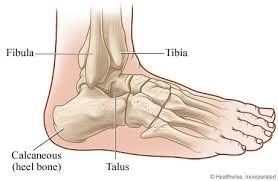
Bones and joints
The ankle joint is formed by the connection of three bones. The ankle bone is called the talus. The top of the talus fits inside a socket that is formed by the lower end of the tibia (shinbone) and the fibula (the small bone of the lower leg). The bottom of the talus sits on the heelbone, called the calcaneus.
Inside the joint, the bones are covered with a slick material called articular cartilage (helps with shock absorption). Articular cartilage is the material that allows the bones to move smoothly against one another in the joints of the body.
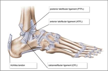
Ligaments and tendons
Ligaments are the soft tissues that attach bones to bones and tendons attach muscles to bones. Both of these structures are made up of small fibres of a material called collagen. The collagen fibres are bundled together to form a rope-like structure. Thickness of the ligament or tendon determines its strength.
Ligaments on both sides of the ankle joint help hold the bones together. Three ligaments make up the lateral ligament complex on the side of the ankle. These include the anterior talofibular ligament (ATFL), the calcaneofibular ligament (CFL), and the posterior talofibular ligament (PTFL).
A thick ligament, called the deltoid ligament, supports the medial ankle (the side closest to your other ankle).
Ligaments also support the lower end of the leg where it forms a hinge for the ankle. Three main ligaments support this area. The ligament crossing just above the front of the ankle and connecting the tibia to the fibula is called the anterior inferior tibiofibular ligament (AITFL).
The posterior fibular ligaments attach across the back of the tibia and fibula. These ligaments include the posterior inferior tibiofibular ligament (PITFL) and the transverse ligament.
'We bring you the latest Garden Route, Hessequa, Karoo news'